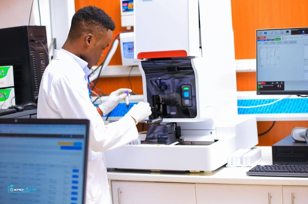
HEMATOLOGY TESTS AVAILABLE
Complete Blood Count (CBC)
The CBC is used as a broad screening test to check for such disorders as anaemia, infection, and many other diseases. It is actually a panel of tests that examines different parts of the blood and includes the following:
Blood Film
A blood film allows the evaluation of white blood cells (WBCs, leucocytes), red blood cells (RBCs, erythrocytes), and platelets (thrombocytes). These cell are produced and mature in the bone marrow and are released into the bloodstream when needed. WBCs’ main function is to fight infection, while RBCs carry oxygen to the whole of the body. Platelets appear as small cell fragments and, when activated, stick together to form a plug as one of the first steps to stop bleeding. The number and type of each cell present in the blood changes but is normally maintained by the body within specific ranges. Values can change at times of illness or stress; intense exercise or smoking can also affect cell counts.
A blood film is a snapshot of the cells that are present in the blood at the time that the sample is obtained. To produce a blood film, a single drop of blood is spread in a thin layer across a glass slide, dried, and then stained with a special dye. Once the stain has dried the slide is looked at under a microscope by a healthcare scientist or haematologist.
HOW IS IT USED
A blood film was once prepared on nearly everyone who had a full blood count (FBC). With the automated blood cell counting instruments currently used, an automated differential is also provided. However, if the presence of abnormal WBCs, RBCs or platelets is suspected, a blood film, examined by a trained eye, is still the best method for identifying immature and abnormal cells.
There are many diseases, disorders and deficiencies that can have an effect on the number and type of blood cells produced, their function and their lifespan. Although usually only normal mature cells are released into the bloodstream, circumstances can force the bone marrow to release immature and/or malformed cells into the blood. When a significant number of abnormal cells are present, they can indicate disease and prompt the doctor to do further testing.
WHEN IS IT REQUESTED?
The blood film is primarily used when a FBC with differential, performed with an automated blood cell counter, shows the presence of abnormal or immature cells. It may also be used when a doctor suspects a deficiency, disease or disorder that is affecting blood cell production, such as an anaemia, decreased or abnormal production of cells in the bone marrow, or increased cell destruction. A blood film may also be requested when a patient is being treated or monitored for a blood cell-related disease.
WHAT DOES THE TEST MEAN?
Findings from the blood film test do not always give a diagnosis but can provide information indicating the presence of an underlying condition and its severity and the need for further diagnostic testing. Blood film findings may include:
RBC (Red blood cells)
Normal, mature red blood cells are uniform in size (7 µm). Unlike most other cells, they do not have a nucleus. They are round and flattened like a doughnut but with a depression in the middle instead of a hole (biconcave). With routine staining, due to the haemoglobin inside the RBCs, they appear pink to red in colour with a pale centre. While not every RBC will be perfect, the presence of many cells that are different in shape or size may indicate a more severe problem. There may be one or more irregularities present and may include:
See the section below for Details on Red Blood Cell Irregularities.
WBC (White Blood Cells)
White blood cells have a nucleus surrounded by cytoplasm. All WBCs come from bone marrow stem cells. In the marrow, they change into two groups: myeloid and lymphoid cells. They then mature into five different types of WBCs.
See the section below for more Details on White Blood Cells.
Platelets
These are cell fragments that come from large bone marrow cells called megakaryocytes. Upon release from the bone marrow, they appear as fragments in the peripheral blood. When there is blood vessel injury or other bleeding, the platelets become activated and begin to clump together to form aggregates. This is the first step in making a blood clot. You must have a sufficient number of platelets to control bleeding. If there are too few, the ability to form a clot is impaired and can be life-threatening. In some people, too many platelets may be produced, which interferes with the flow of blood and increases a person's risk of developing a blood clot. These same people may also experience bleeding because many of the extra platelets may be dysfunctional even though they appear normal.
Details of platelet number and size is usually part of a FBC. An abnormally low number or high number of platelets may be further evaluated by preparing a peripheral blood film to visualise any anomalies in shape or size directly.
Platelet Function Tests
Platelets are small cell fragments and are found in the blood along with red cells and white cells. Platelets are produced in the bone marrow and released into the blood where they play an important role in coagulation (blood clotting), helping to stop bleeding when blood vessels are injured. These small, disc-shaped cells usually live for around 5-10 days in the blood before they are destroyed.
Platelets are very important for efficient blood coagulation and preventing unnecessary or excessive blood loss. If there are too few platelets, or if the platelets that are present don’t function properly, then there may be problems with blood clot formation. Platelet function tests are used to indirectly evaluate how well a person's platelets work in helping to stop bleeding within the body.
When there is an injury to a blood vessel and bleeding begins, platelets help to stop bleeding in three ways. They:
These reactions result in the formation of a loose platelet plug in a process called primary haemostasis. At the same time, activated platelets support the coagulation cascade, a series of steps that involves the sequential activation of proteins called clotting factors. This is called secondary haemostasis and the two processes result in the formation of a stable clot that remains in place until the injury has healed.
If there are insufficient platelets or if they are not functioning normally in any of the three main ways, a stable clot may not form and a person may be at an increased risk of excessive bleeding. The number of platelets in blood can be determined with a platelet count and can help diagnose disorders having to do with too many or too few platelets. However, the platelet count simply provides information on the number of these cells that are present in blood; it does not reveal if these cells are functioning properly. Specialised tests, called the platelet function tests, are used when there is suspicion of a platelet function defect.
Platelet function tests involve testing the response of an individual's platelets to a variety of stimuli (platelet agonists) in the laboratory, using the timing and magnitude of platelet aggregation to the platelet agonists to measure the ability of platelets to promote clotting in a sample of blood. There are a variety of tests available but no single test that identifies all problems with platelet function. Also, there is no widespread agreement on which test(s) is best for each circumstance.
In addition to evaluating people for excessive bleeding, platelet function tests may be used in other situations. There are situations in which it is desirable to decrease the ability of platelets to aggregate, as in for people who are at an increased risk of developing a dangerous blood clot or at increased risk for heart attacks. These people may be prescribed medications that reduce platelet activation or reduce their ability to aggregate. People on these types of anti-platelet medications, such as low-dose aspirin or clopidogrel, may have platelet function tests done as a way of monitoring their treatment. However, there is currently no consensus among medical experts on the usefulness of platelet function tests in anti-platelet therapy.
D-dimer
When a vein or artery is injured and begins to leak blood, a sequence of clotting steps and factors (called the coagulation cascade) is activated by the body to limit bleeding and create a blood clot to plug the hole. During this process, threads of a protein called fibrin are produced. These threads are cross-linked (glued together by a protein called thrombin) to form a fibrin net that catches platelets and helps hold the forming blood clot together at the site of the injury.
A positive D-dimer indicates the presence of an abnormally high level of cross-linked fibrin degradation products in your body. It tells your doctor that there has been significant clot (thrombus) formation and breakdown in the body, but it does not identify the location or cause. An elevated D-dimer may be due to a VTE or but it may also be due to a recent surgery, or trauma, infection, liver or kidney disease, cancers, in normal pregnancy but also some diseases of pregnancy such as eclampsia.
A normal D-dimer test means that it is unlikely you have an acute blood clot or disease causing abnormal clot formation and breakdown. Most doctors agree that a negative D-dimer is most valid and useful when the test is done on patients that are considered to be low-risk. The test is used to help exclude a clot as the cause of symptoms.
D-dimer is recommended as an ‘additional test’. It should not be the only test used to diagnose a disease or condition. Both increased and normal D-dimer levels may require follow-up and can lead to further testing.
Erythrocyte Sedimentation Rate (ESR)
ESR is an indirect measure of the degree of inflammation present in the body. It actually measures the rate of fall (sedimentation) of erythrocytes (red blood cells) in a tall, thin tube of blood. Results are reported as how many millimetres of clear plasma are present at the top of the column after one hour. Normally, red cells fall slowly, leaving little clear plasma. Increased blood levels of certain proteins (such as fibrinogen or immunoglobulins, which are increased in inflammation) cause the red blood cells to fall more rapidly, increasing the ESR.
WHAT DOES THE TEST RESULT MEAN?
Doctors do not base their decisions solely on ESR results. You can have a normal result and still have a problem.
A very high ESR usually has an obvious cause, such as an infection. The doctor will use other follow-up tests, such as cultures, depending on the patient’s symptoms.
Moderately elevated ESR occurs with inflammation, but also with anaemia, infection, pregnancy, and old age.
A rising ESR can mean an increase in inflammation or a poor response to a therapy; a decreasing ESR can mean a good response.
A common cause of high ESR is anaemia, especially if it is associated with changes in the shape of the red cells; however, some changes in red cell shape (such as sickle cells in sickle cell anaemia) lower ESR. Kidney failure will also increase ESR. People with multiple myeloma or Waldenstrom’s macroglobulinaemia (tumours that make large amounts of immunoglobulins) typically have very high ESR even if they don't have inflammation.
Although a low ESR is not usually important, it can be seen with polycythaemia (a condition where a patient makes too many red blood cells), with extreme leucocytosis (patient has too many white blood cells), and with some protein abnormalities.
CD4 and CD8
CD4 and CD8 cells are lymphocytes that have markers on the surfaces of the cells called CD4 and CD8. They are types of white blood cells that fight infection, and they play an important role in your immune system function. CD4 and CD8 cells are made in the bone marrow, and mature in the thymus gland, a small gland found in the upper chest. They circulate throughout the body in the bloodstream, spleen and the lymph nodes.
CD4 cells are sometimes called T-helper cells. They help to identify and trigger the attack and destruction of specific
The test result for PT depends on the method used; results will be measured in seconds. An increased Prothrombin time or INR means that your blood is taking longer to form a clot. If you are not taking anticoagulant drugs and your PT is prolonged, additional testing may be necessary to determine the cause.
For monitoring of vitamin K-antagonists, such as warfarin, PT results are adjusted to the International Normalised Ratio (INR). People on anticoagulant drugs usually have a target INR of 2.0 to 3.0 (i.e. a prothrombin time 2 to 3 times as long as in a person not on warfarin with normal clotting, using standardised conditions). For some people who have a high risk of clot formation, the INR needs to be higher: about 3.0 to 4.0. Your healthcare team will use the INR to adjust your drug to get the PT into the range that is right for you.
The PT is often performed along with another clotting test called the aPTT (or sometimes the PTT or KCCT). Comparison of the two results can give your healthcare team information as to the cause of a bleeding problem.
| PT RESULT | APTT RESULT | POSSIBLE CONDITIONS PRESENT |
|---|---|---|
| Prolonged | Normal | Liver disease, decreased vitamin K, decreased or defective factor VII |
| Normal | Prolonged | Decreased or defective factor VIII, IX, XI or XII, von Willebrand disease, or lupus anticoagulant present |
| Prolonged | Prolonged | Decreased or defective factor I, II, V or X, liver disease, disseminated intravascular coagulation (DIC) |
| Normal | Normal | Decreased platelet function, thrombocytopenia, factor XIII deficiency, mild deficiencies in other factors, mild form of von Willebrand’s disease, weak collagen |
